Senior Thesis Student Projects - 2013
- Courtney Smith
Solubility, Antibacterial Properties and Cytocompatibility of Silver Coated Glass Microspheres.
- Abstract: Amber glass particles (<90 μm) were used to synthesize glass microspheres which were then coated with silver (Ag). These silver-coated glass microspheres were compared against the uncoated analogous microspheres. The hypothesis of this experiment was that the silver-coated microspheres would eliminate bacteria in anti-bacterial testing, be non-cytotoxic and also alter the solubility of the spheres, compared to control uncoated microspheres. Both Ag coated and uncoated microspheres were characterized using X-ray diffraction (XRD), scanning electron microscopy (SEM) and energy dispersive X-ray analysis (EDS), optical microscopy, advanced surface area and porosity (ASAP). Inductively coupled plasma atomic emission spectroscopy (ICP-OES) was used to determine the solubility, while antibacterial and cell viability testing were used to determine the therapeutic effect. XRD analysis indicated no crystallinity in the initial powder stage or in the uncoated microsphere stage, but showed crystallinity of silver peaks in the silver-coated glass microspheres. SEM determined that the silver coating remained at the surface of the spheres and that there was little diffusion through the bulk. Antibacterial testing proved that the silver-coated glass microspheres inhibited the E. coli. Neither the silver-coated nor the uncoated microspheres were deemed cytotoxic and the ion release profiles indicate an increase in ion release when the glass microspheres are coated in silver.
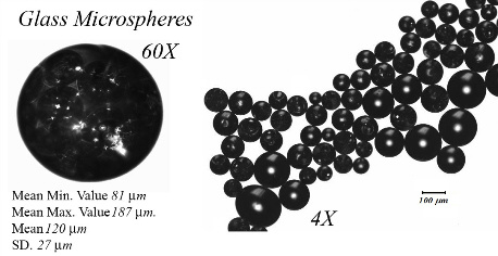
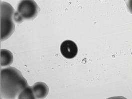
- Glass Microspheres Images taken with Olympus Optical Microscope.
- Caelen Clark
Characterization of Anodized Titanium Substrates for Orthopaedic Applications.
- Abstract: Titanium and its alloys are considered to be the best choice for the replacement of load-bearing hard tissue. Due to the passive oxide layer that forms on the surface of titanium it is considered to be a biocompatible material. Surface modifications are required for the promotion of the osseointegration in these materials. Anodic oxidation is a simple method of surface modification that can be used to increase the osseointegration of titanium implants. Anodic oxidation can result in the formation of a layer of TiO2 nanotubes on the metal surface. This work seeks to characterize the surface morphology and biocompatibility of anodized titanium. Atomic force microscopy, scanning electron microscopy and scanning laser profilometry were used to image the surface of anodized titanium samples. Additionally, the surface roughness of the samples was analyzed using laser profilomerty. To evaluate the biocompatibility of the samples MC3T3 osteoblast cells were seeded onto the titanium samples. The morphology and spreading of the cells was evaluated using SEM.
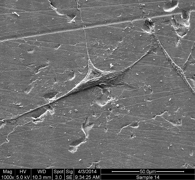
- Osteoblast Cell Adhered to the Surface of the Titanium Substrate.
- Matthew Strohmayer
Preliminary Study of Polymer-Nanocomposite Synthesis, Simulated Body Fluid and Mechanical Testing.
- Abstract: Nano materials are currently being incorporated into polymer composites to improve properties such as weight, strength and bioactivity. In this study, carbon nanotubes (CNT) and Si Zr nano hydroxyapatite (nHAP) were added to Ethoxylated Bisphenol A Dimethacrylate (EBPADMA). It was found after initial synthesis of the samples that a thickening agent, fumed silica (FS), was required to prevent the pre-polymerized polymer from flowing too much. It was also found that a combination of FS and sonication promoted the nanotubes to disassociate and remain suspended in solution. Four sets of samples were synthesized, the control being just EBPADMA, the second EBPADMA and FS, the third EBPADMA FS and CNT, the final being comprised of EBPADMA FS CNT and nHAP. Then simulated body fluid (SBF) was made and five samples of disks and cylinders were immersed in 10 ml for 1, 7, and 30 days. After each time period, the mechanical strength was tested. It was found that the nano composites had a lower compressive strength between each sample and had a similar strength across the time periods, although the average strength dropped the standard deviations overlap. In biaxial flexural strength (BFS), the samples showed a reduction form the control and a slight decrease with the first two time periods and similar numbers on the final 30 day test. It was also found that after 30 day that the average BFS of the samples was higher than the control. Samples were also immersed in 60 ml of SBF and the calcium phosphate (CaP) deposition was looked at with SEM and EDS after 1, 7, and 30 days. All one day samples showed next to no CaP deposition under the SEM. The 7 day samples were seen to have some deposition, but not a significant amount. The 30 day samples had a significant 2 amount to the point where the deposition was clearly visible. The identity of this was confirmed by EDS. Based on this information, this polymer nanocomposite has potential application as bone cements.
- Scanning Electron Microscopy Image of Nano-composite Material.
- Lydia Boutelle
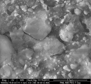
Characterization of Silver Coated Bioactive Glass Particles and Glass-Polymer Hydrogel Composites.
- Abstract: Studies have shown that the addition of the silver ion (Ag+) to bioactive materials results in improved antimicrobial properties. The distribution of silver-coated bioactive glass particles throughout a two-phase hydrogel, consisting of synthesized carboxymethyl cellulose-alpha (CMC-α) and dextran-β, was studied to determine the potential for these materials to act as a sterilizing composite used for bone void filling. Surface area analysis determined that silver-coated particles had an average surface area of 0.1366 m2/g and the uncoated particles had an average surface area of 0.2149 m2/g. Elemental analysis of glass particles through Energy Dispersion Spectroscopy determined the elemental composition of each glass and images of microstructure were captured using Scanning Electron Microscopy. The inhibition zones of silver-coated glass particles in gel were compared to particles of uncoated bioactive glass in gel, and to that of control hydrogels, which contain no glass particles, swabbed with Escherichia coli. Antibacterial testing determined that Ag-coated gel composites exhibited the largest inhibition zones compared to uncoated gel composites. Broth dilution tests were conducted to observe the absorbance of each gel. A control gel, Ag-coated gel, uncoated gel, and bacteria broth were diluted with e. coli, and compared with a sterile broth. Dilution tests determined that Ag-coated gels exhibited the second-highest absorbance levels and closest to sterile broth absorbance levels. As bacterial resistance to current antibiotics increases, and the level of new antibiotics in development dwindles, studies such as these can further aid in the progression of silver use in wound healing, and antibiotic-resistant bacterial applications.
- Scanning Electron Microscopy of Freeze dried Hydrogel with Salt Deposits on the Surface.
- Nicole Keenan
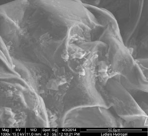
Synthesis and Characterization of Gallium-Containing Bioactive Glass-Polymer Hydrogel Composites.
- Abstract: According to the National Cancer Institute, only 10-40% of patients who experience local recurrence of cancer within bone survive. Due to its previously determined heightened bioactivity and effectiveness against several forms of cancer, gallium has been hypothesized to be a useful component of bone void fillers by potentially decreasing the rate of bone turnover and preventing the return of cancer cells post-resection. Furthermore, hydrogels are being analyzed as the delivery mechanism for these Ga-containing bioactive glasses. Several advantages, such as a non-thermal setting reaction and the lack of harmful byproducts, are what make hydrogels an attractive candidate for this application. The goals of this study were to synthesize uniform hydrogels to act as a carrier for Ga-containing bioactive glass particles, and to examine the interactions between the hydrogels and bone tissue. These glass-hydrogel composites were assessed based upon pH, swelling ratios, rehydration tendencies, and polymer-to-water ratios, while information pertaining to their interaction with bone was assessed using scanning electron microscopy (SEM) and energy dispersive x-ray spectroscopy (EDS). It was determined that glass particles were moderately disbursed throughout the hydrogels, and that these glass-hydrogel composites effectively attach to bone. Future studies include tailoring the properties of the hydrogels and adjusting the added glass content in order to achieve optimal suspension of glass particles.
- Hydrated Hydrogel and Freeze Dried Hydrogel.
- Scott De-Franco Norton
Bioabsorbable Coronary Stents for the Reduction of Thrombosis and Restenosis.
- Abstract: Three separate magnesium based sample were compared together to conclude the ideal material to be used in coronary stenting. The samples of Mg, Mg-Zn, and Mg-Y-Zn were put through a variety of microstructural, compression and bacterial testing to compare with one another. Agglomeration of particles increased as dopants were added to the Mg and ended in the overall mechanical and antibacterial failure of Mg-Y-Zn. Mg-Zn withstood a high compression test as well as showed a homogenous sample surface after sintering process. The Mg-Zn showed good antibacterial properties when incubated with Escherichia coli. The hypothesis that adding dopants to Mg would increase the mechanical and biocompatible properties of the material was disproven; but it was positive to find that Mg-Zn had test results which comply with being an ideal material which can now be used in further analysis and testing.
- Mg-Zn-Y ceramic Discs produced for Mechanical Testing.
- Heather Liggett
Investigating Osteoblast Cell Populations Using Cyclic Voltammetry.
- Abstract: In this experiment, the metabolic activity of viable osteoblast MC3T3 cells is analyzed using cyclic voltammetry, and its results are correlated to the findings of an automated trypan blue exclusion assay to determine the feasibility of cyclic voltammetry as a novel method for cell viability analysis. It is hypothesized that an increasing population of cells will yield greater concentrations of metabolites, therefore leading to observed electrochemical behaviors that change as a function of cell population. Osteoblast cells were cultured, imaged with optical light microscopy, and seeded onto a 12-well cell culture plate in increasing concentrations that were determined using a haemocytometer. The viability of cell populations were analyzed after incubation using the trypan blue assay and cyclic voltammetry. The results of cyclic voltammetry testing determined that an inverse relationship exists between the concentration of osteoblast cell metabolites in solution and the characteristic electrochemical behaviors observed. It is concluded that in comparison with currently utilized cell viability assays, cyclic voltammetry is strictly qualitative for the purposes of this experiment, but may be further developed into a simple, quick, and cost effective quantitative method with the application of significant additional research.

- Sufrace adheered osteoblasts at a concentration of 100k/ml.
- Peter Saunders
Synthesis of PLGA Microspheres with Encapsulated Protein -Lysozyme.
- Abstract: Traditional drug delivery methods tend to be inefficient and cannot provide site-specific drug delivery or controlled drug concentration once subcutaneously injected. Over the years Poly(lactic-co-glycolic acid) has emerged as a polymer that can be used to provide control to variables that made drug delivery systems inefficient in the past. Due to these added benefits as well PLGA’s biocompatibility with the human body research was conducted to synthesize and perform fundamental analysis on PLGA microspheres with encapsulated lysozyme. In particular, research was performed to determine the encapsulation efficiency and drug load of technique used to synthesize microspheres along with factors that affect sphere stability. In addition to the laboratory research performed, in-depth review of peer-reviewed scientific literature was completed to obtain a better understanding of the properties of PLGA and they can be modified.
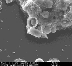
- Scanning electron microscopy of PLGA microspheres at 1000X.
- Kelly-Jo Beck
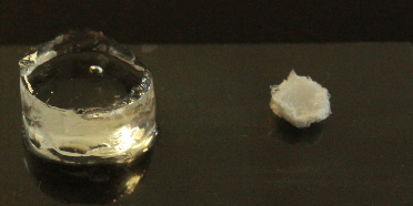
Characterization of TiO2 containing glass-ceramic scaffolds.
- Abstract: Bioactive glass based scaffolds are currently being investigated for applications in bone tissue engineering. This study was conducted to determine the effect TiO2 will have on the structure of bioengineering scaffolds at varying concentrations. XRD was undergone to confirm the amorphous properties of the five glass-ceramic samples, while EDS was done to confirm the absence of Ti in the control sample. PSA was used to compare the particle size of each sample, as smaller particle size is optimal for sintering and fabrication of scaffolds. Images were taken using a scanning electron microscope before scaffold production to also compare particle size. A polymer foam replication technique was used to prepare porous scaffolds of varying compositions. After scaffolds were formed for the control sample and the scaffold with the highest TiO2 concentration, SEM images were taken to examine the structure and macro- and micro-porosity of the scaffolds. It was determined that the addition TiO2 to the powders improved the structure of the scaffolds by forming a more defined structure and increasing the macro- and micro-porosity of the scaffold.
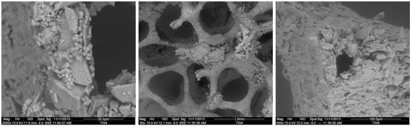
- Heat treated glass ceramic scaffolds with the polymer burnout from a strut on the far right.
Senior Thesis Student Projects - 2012
- Laura Haas
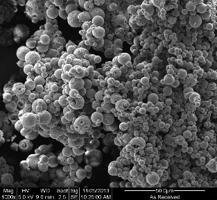
Characterization and antibacterial efficacy of silver coated glass microspheres.
- Abstract: Amber glass particles of varying size (50 to 100 μm) were synthesized and coated with silver (Ag) to produce silver-coated particles. These were compared against the uncoated analogous particles. The silver-coated microspheres will kill the bacteria in anti-bacterial testing, whereas the uncoated microspheres will remain the same was the hypothesis for this experiment. Each glass and silver-coated glass was characterized using X-ray diffraction, particle size analysis, advanced surface area and porosity, scanning electron microscopy and energy dispersive x-ray analysis, and antibacterial testing. X-ray diffraction analysis indicated no crystallinity in the initial powder stage and the uncoated glass stage, but showed crystallinity of silver peaks once the glass particles were coated. Surface area analysis determined that the silver coating of the glass particles resulted in a decrease in surface area from 0.0895 to 0.065 m2/g. Scanning electron microscopy determined that the silver coating remained at the surface and that there was little diffusion through the bulk. Antibacterial (Escherichia coli- 13 mm) testing determined that the silver-coated glass microspheres exhibited the largest inhibition zones compared to the uncoated glass microspheres. The uncoated glass microspheres were engulfed by the bacteria and proved the hypothesis to be true.

- Silver coated microspheres antibacterials efficacy against E. coli on a plate (left) and individual sphere (right).
- Emily Allen
Bioactivity and solubility of gallium containing glass polyalkenoate cements.
- Abstract: In current orthopedic applications, bone cements are usually comprised of polymethylmethaccrylate (PMMA), which is used to fill voids within the skeleton following trauma or surgery. In the present market glass polyalkenoate cements (GPCs) are used in dentistry, and have begun to be used as a bioactive bone cement with proven efficacy in bone regeneration and therapeutic effects. As these bioactive glasses are beginning use in medical settings, the current objective of this work is to determine the bioactivity of gallium based polyalkenoate cement. Three glasses were formulated for this study: a gallium free control (LCon) composed of CaO-ZnO-SiO2; and two glasses (LGa-1, LGa-2) which had increasing amounts of gallium at the expense of zinc. These glasses were annealed and subjected to particle size analysis and X-ray diffraction prior to disk formation. The GPC disks were then made in pairs using a 1g-glass powder, 0.375g E11 PAA, and 0.375g deionized water composition. Once set these disks were placed into simulated body fluid (SBF) for testing times of 1, 7, and 14 days. After the disks were extracted from the simulated body fluid after the given testing times they were subjected to SEM and EDX analysis. Additional testing of antibacterial properties and bioactivity should be done before the clinical application of this GPC can be used for orthopedic applications.
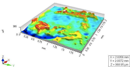
- Laser profilometry of GPCs surface after 14 days immersion in Simulated Body Fluid.
- Doughlas Cohen *
Synthesis of biodegradable PLGA microspheres with encapsulated bovine serum albumin.
- Abstract: Conventional drug delivery mechanisms can be inefficient with regards to dosage and tissue specificity as they rely on a dispersive approach to treat the affected area. A more targeted and controlled methodology may be beneficial as one could effectively control the dosage to treat the same medical conditions. For this reason, research was conducted to synthesize and characterize biodegradable Poly(lactic-co-glycolic acid) microspheres with encapsulated bovine serum albumin, to demonstrate their potential as a biocompatible drug transporter. More specifically, this work was conducted to test the effectiveness of the utilized synthesis technique, and to gain insight on the PLGA microsphere protein encapsulation process. Through successive trials, microspheres were successfully synthesized via a double emulsification process – with the BSA dispersion and subsequent encapsulation aided by a Branson Sonfier 450. The resulting microspheres were then rapidly converted to a cryogenic state by way of liquid nitrogen cooling and then freeze dried using Labconco TriadTM for 24 hours under high vacuum. Once freeze dried the microspheres were stored at -80°C in a Fisher Scientific Isotemp freezer. Particle size analysis from 2-60 μm was conducted utilizing a Beckman Coulter Multisizer 4; with 90% of the microspheres recorded to have an average diameter less than or equal to 4.4 μm, and an average surface area of 12442.5 cm2/mL. The microspheres were also visually analyzed using reverse transmission optical microscopy and scanning electron microscopy. SEM images revealed well defined PLGA microspheres as well as some nanospheres, and EDS analysis confirmed their chemical purity.
- SEM image of clustered PLGA microspheres.
- Tracie McGinnity *
Investigating the structure and solubility of SiO2-CaO-ZnO-Na2O-Ga2O3 glasses.
- Abstract: This study focuses on determining any structural changes that occur in a series of bioactive glasses in which Zinc (Zn) has been incrementally replaced by Gallium (Ga). Structural analysis was conducted using Differential Thermal Analysis (DTA), which yielded a thermal profile of each glass. Also included in the study was whether or not the incorporation of Ga affects the formation of a calcium phosphate layer on the glass surfaces after being submerged in simulated body fluid (SBF). These glasses included Ga because of its antibacterial properties and anti-cancer abilities in vivo.
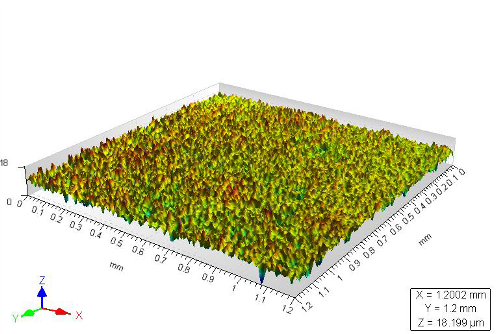
- Laser profilometry of glass surface after 14 days immersion in Simulated Body Fluid.
- Matthew Elmer
Fabrication of torlorn/carbon nanotube composite polymers.
- Abstract: Polymer matrix composites (PMC) are found in automotive and aerospace applications where a material is desired to have high strength and stiffness with minimal weight penalty. Contemporary materials science research involving PMCs has emphasized the use nanoscale reinforcements to see significant property improvements. The aim of this study is to fabricate bulk nanocomposites from Torlon 4000TF poly-amide-co-imide (PAI) and carbon nanotubes (CNT). Paramount to this goal is achieving stable dispersions of nanoparticles in order to form a homogenous final part. The thermal and mechanical benefits of the reinforcing phase are optimized in this situation. A noncovalent functionalizer branded Kentera is used to form strong interfacial region by exploiting a pi-pi bonding network. Several processing methods are attempted to identify the most feasible way to integrate CNTs to the matrix. There are two primary tactics to the processability study; solution mixing and non-solution mixing. The general approach of solution mixing starts with dissolving the Torlon and dispersing nanoparticles in similar solvents. Next the solvent is to be extracted in a way that does not interfere with the even distribution both phases. Vacuum distillation, anti-solvent and Soxhelt extraction, and freeze-drying methods are classified solution mixing technique. Alternatively the non-solution mixing entails dispersing both Torlon and nanoparticles in an easily volatile medium. This method is known as the sonic hot water bath. After addition of the nanoparticles samples were formed via compression molding. The composites are characterized with differential scanning calorimetry (DSC), Fourier transform infrared spectroscopy (FT-IR), scanning electron microscopy (SEM), energy dispersive X-ray spectroscopy (EDX), and ASTM standard D5405 measuring fracture toughness. Addition of CNTs causes no mechanical enhancement, however this is possible more attributable to issues with compression molding. Average critical stress intensity factor for fracture toughness of 1.99MPa√m (as-received), 1.14MPa√m (sonic hot water bath), and 0.51MPa√m (vacuum distillation) were measured. It was concluded that vacuum distillation remains the most suitable process with refinement of the molding procedure. This is because of a 25ºC increase in the glass transition over raw materials and success of dispersing nanomaterials.
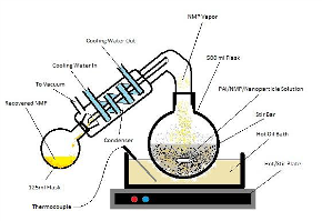
- Solvent extraction process sued in fabricating composite materials.
- James Whyel
Characterization of hydroxyapatite particles.
- Abstract: Hydroxyapatite is one of few materials to be classified as bioactive. It is similar to the mineral component of bones and hard tissues found in mammals. Bioactive materials have potential in practically every medical procedure, from cancer treatment to bone replacement. Hydroxyapatite (HA) is becoming increasingly attractive for bone cements and bone implants, but in order to harness the full potential of this compound, further studies are needed regarding its bioactivity and strength upon incorporation in a cement. Wear debris caused from wear abrasion and lack of strength can result in inflammation, bone loss, and overall failure of an implant. In order for bone cements to be used as implants, the material must include contrast agents that allow for their visual detection in radiographic examinations. It is believed that incorporating a bioactive component like HA, along with proper contrast agents, could tackle the clinical need for increased strength, toughness, and wear resistance of bone cements. The samples analyzed in this study have the potential to produce bone cements that possess improved mechanical properties, increased bioactivity, improved adhesion and bone growth, and enhanced stability when dispersed in the polymer precursors. Regarding this study, bioactive particles were prepared by an outside source and were characterized using a number of techniques, including SEM, EDX, XRD, BET surface area, and SBF analysis. This study was undertaken in order to characterize bioactive dopants that can improve the mechanical properties, biological response and visualization of orthopaedic bone cements.
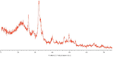
- X-ray diffraction of hydroxyapatite nanoparticle.
* Oustanding Research Award presented by Sigma Xi, the research honor society.